
Dental X-ray
Digital intraoral, panoramic, cephalometric and CBCT 3D imaging — low dose, on-screen in seconds, kept on file.
- Comprehensive Assessment
- Digital Imaging When Needed
- Personalized Treatment Plan
- Transparent Recommendations
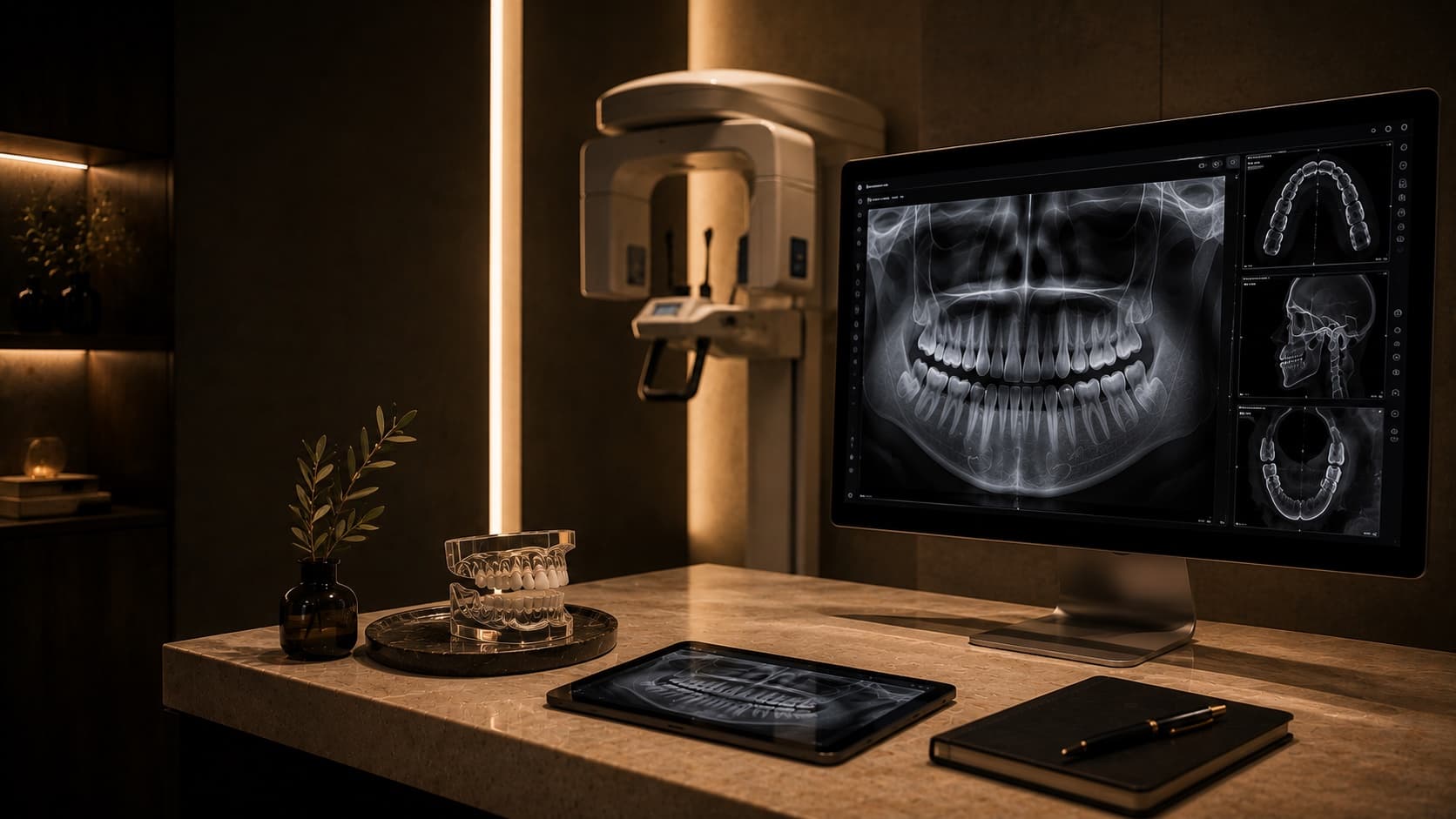
When imaging is needed.
X-rays show what an exam cannot — decay between teeth, bone levels under the gum, root structure, impacted teeth and jaw anatomy. We take imaging only when it changes what we will do, and we choose the smallest scan that answers the clinical question.
- Routine check-up to screen for decay between teeth
- Toothache or sensitivity with no visible cause
- Before a root canal, extraction or implant
- Orthodontic assessment and planning
- Wisdom tooth evaluation
- Tracking a child's developing teeth and bite
Four imaging modalities.
Tap a treatment for what it's used for, how it's planned, and how it's kept comfortable.
- Best for
- Routine check-ups, toothache, deep fillings, suspected fracture, pre-extraction
- Planning
- Indicated by clinical findings; one to four films in most sessions
- Comfort
- Small sensor placed inside the mouth; image captured in a fraction of a second
Ask about digital intraoral x-raysPatient benefits- High-detail view of a single tooth or area.
- Lower dose than older film X-rays.
- Image visible on-screen in seconds.
- Image enlarged and adjusted for clarity.
- Best for
- General check-up overview, wisdom teeth, orthodontic planning, jaw assessment
- Planning
- Stand or sit; head positioned in the machine; no intraoral sensor
- Comfort
- Non-invasive; no sensor in the mouth; 10–20 seconds
Ask about panoramic opg imagingPatient benefits- Whole-mouth overview in a single image.
- No sensor inside the mouth — comfortable for children and anxious patients.
- Captures wisdom teeth, sinuses and jaw joints.
- Lower dose than older film systems.
- Best for
- Braces and aligner planning, jaw growth assessment, orthognathic surgical planning
- Planning
- Standard pairing with OPG at orthodontic consultation
- Comfort
- Non-invasive; head supported with chin rest; a few seconds
Ask about cephalometric x-rayPatient benefits- Objective measurement of jaw and facial proportions.
- Predicts growth patterns in children and teens.
- Standard input for orthodontic and surgical planning.
- Comparable year over year.
- Best for
- Implant planning, impacted wisdom teeth, complex root canals, jaw pathology, TMJ assessment
- Planning
- Justified by clinical reason; focused-field scan to limit dose
- Comfort
- Non-invasive; open machine (not a tunnel); under 20 seconds
Ask about cbct 3d imagingPatient benefits- Measures bone width, density and nerve position before implant surgery.
- Reveals hidden canals and root anatomy missed on 2D.
- Plans wisdom tooth extractions near sensitive nerves.
- Assesses jaw joints and airway in TMJ and sleep cases.
Planned with precision. Performed with care.
We image to a question, not to a checklist. A toothache may need one bitewing; an implant case needs CBCT; orthodontic planning needs an OPG and ceph. We keep your imaging in your file so we can compare year over year, and we share scans with your other specialists when needed.
Justified, not routine
Every X-ray is justified by a clinical reason. We do not duplicate scans you already have if they are recent and adequate.
Low-dose digital
Digital sensors use a fraction of the radiation of older film systems. CBCT uses focused-field protocols to limit dose to the area being studied.
Lead apron and shielding
Standard for every patient, every time. Thyroid collars are offered where the scan field allows it.
Instant, shared on-screen
Images appear in seconds on a chairside monitor so we can show you what we see and explain the plan with the scan in front of you.
What to expect, step by step.
- 01
Clinical exam first — imaging is requested only if it will change the plan.
- 02
Lead apron and thyroid collar applied.
- 03
Metal items (jewellery, glasses, hairpins) removed for OPG, ceph and CBCT.
- 04
Scan captured — 1–20 seconds depending on modality.
- 05
Image appears on-screen for immediate review.
- 06
Findings discussed with you on the chairside monitor.
- 07
Scan saved to your record for future comparison.
Why digital imaging matters.
A clinical exam tells us about the surface of the teeth. Imaging shows what is happening below the gum, between teeth and inside bone. Digital sensors keep the dose low, the result fast, and the file ready to share.
- Catches decay, infection and bone loss before symptoms appear.
- Plans implants, surgery and root canals with measured anatomy.
- Shows you what we see so treatment decisions are clear.
- Replaces guesswork with measurement.
- Lower radiation than older film systems, with shielding for every patient.
Not sure which treatment you need? Talk to a specialist on WhatsApp →
Common questions.
Direct answers to the questions patients ask before booking — pain, sedation, recovery, and the steps from consultation to result.
Yes. Digital sensors use a fraction of the radiation of older film X-rays. A single intraoral X-ray exposes you to less radiation than a few hours of background exposure at sea level. We use lead aprons every time and only take films that are clinically justified.
No. Routine bitewings are recommended at intervals based on your risk — every 6–24 months for adults, depending on decay history. We do not repeat scans for the sake of it.
A standard X-ray is a 2D image. A CBCT is a 3D scan that shows depth, width and the relationship between teeth, bone and nerves. We use CBCT for implants, surgery and complex root canals — not for routine check-ups.
No. The CBCT machine is open — you stand or sit, and the arm rotates around your head. There is no tunnel. The scan itself is finished in under 20 seconds.
We usually postpone routine X-rays until after pregnancy. If you have an emergency, infection or significant pain, the risk of leaving it untreated is often higher than the risk of a single low-dose X-ray with full shielding. We will discuss the options before any scan.
A single X-ray is safe for you. The staff member takes multiple X-rays every day across many patients, and stepping out limits their cumulative exposure over years. It is a standard radiation safety protocol worldwide.
Yes. If you have recent and adequate imaging from another clinic, we will use it and avoid duplicating the scan. Bring them on a USB or have them emailed before your appointment.
Yes. Images appear on the chairside monitor immediately. We go through what we see with you before discussing treatment options.
The next step is a conversation.
Send a message, share what's going on, and we'll take it from there.
Prefer to call? +971 50 177 9179