
Endodontics
Microscope-guided root canal therapy — save the natural tooth, with precision.
- Comprehensive Assessment
- Digital Imaging When Needed
- Personalized Treatment Plan
- Transparent Recommendations
When a root canal is the right call.
Root canal treatment is needed when the pulp inside a tooth becomes inflamed or infected — from deep decay, a crack, trauma, or a previous restoration that has begun to leak. Treating it early saves the tooth.
- Persistent tooth pain that lingers after hot or cold
- Pain when biting or chewing
- Swelling or a small bump on the gum near the tooth
- A tooth that has darkened or discoloured
- A deep crack or fracture exposing the pulp
- A previously root-treated tooth that has become painful again
Five endodontic treatments.
Tap a treatment for what it's used for, how it's planned, and how it's kept comfortable.
- Best for
- Inflamed or infected pulp, deep decay reaching the nerve, cracked teeth, trauma
- Planning
- Digital X-ray, microscope-led canal mapping, CBCT for complex anatomy
- Comfort
- Local anesthesia; laughing gas on request
Ask about microscopic root canal treatmentPatient benefits- Higher success rate than conventional root canal.
- Hidden canals and fine fractures are found and treated.
- More natural tooth structure preserved.
- Most cases done in one appointment.
- Lower risk of re-infection over time.
- Best for
- Persistent infection after root canal, blocked canals, cysts at the root tip
- Planning
- CBCT 3D imaging to locate the infected root tip and surrounding anatomy
- Comfort
- Local anesthesia; small incision with fine sutures
Ask about endodontic micro-surgery (apicoectomy)Patient benefits- Saves a tooth that would otherwise be extracted.
- Minimally invasive compared with traditional oral surgery.
- Faster healing — most patients back to normal activity within days.
- Precise removal of only the infected tissue.
- Best for
- Teeth where a previous root canal instrument has separated inside the canal
- Planning
- CBCT to locate the fragment and assess canal curvature before attempting retrieval
- Comfort
- Local anesthesia; performed under microscope with ultrasonic tips
Ask about removal of broken instrumentsPatient benefits- Saves a tooth that would otherwise need extraction.
- Restores full canal access for cleaning and sealing.
- Microscope and ultrasonic approach protects the root.
- Often resolves chronic pain or swelling caused by trapped infection.
- Best for
- Previously root-treated teeth with new pain, swelling, abscess or changes on imaging
- Planning
- CBCT to identify missed canals, persistent infection or coronal leakage
- Comfort
- Local anesthesia; performed in one or two visits depending on complexity
Ask about complex re-treatmentPatient benefits- Avoids extraction and keeps the natural root.
- Addresses the actual cause of failure, not just the symptoms.
- Microscope finds canals that were missed the first time.
- Restores comfort and bite without an implant or bridge.
- Best for
- Heavily broken-down teeth after root canal, especially molars taking heavy bite load
- Planning
- Digital scan and bite analysis to design a core and crown that match your occlusion
- Comfort
- Local anesthesia; usually one to two visits after the root canal is complete
Ask about restorative endodontics (post & core, crown)Patient benefits- Protects the root-treated tooth from vertical fracture.
- Seals the canal from re-infection through the crown.
- Restores full bite force and chewing function.
- Looks like a natural tooth — no visible metal margins.
Planned with precision. Performed with care.
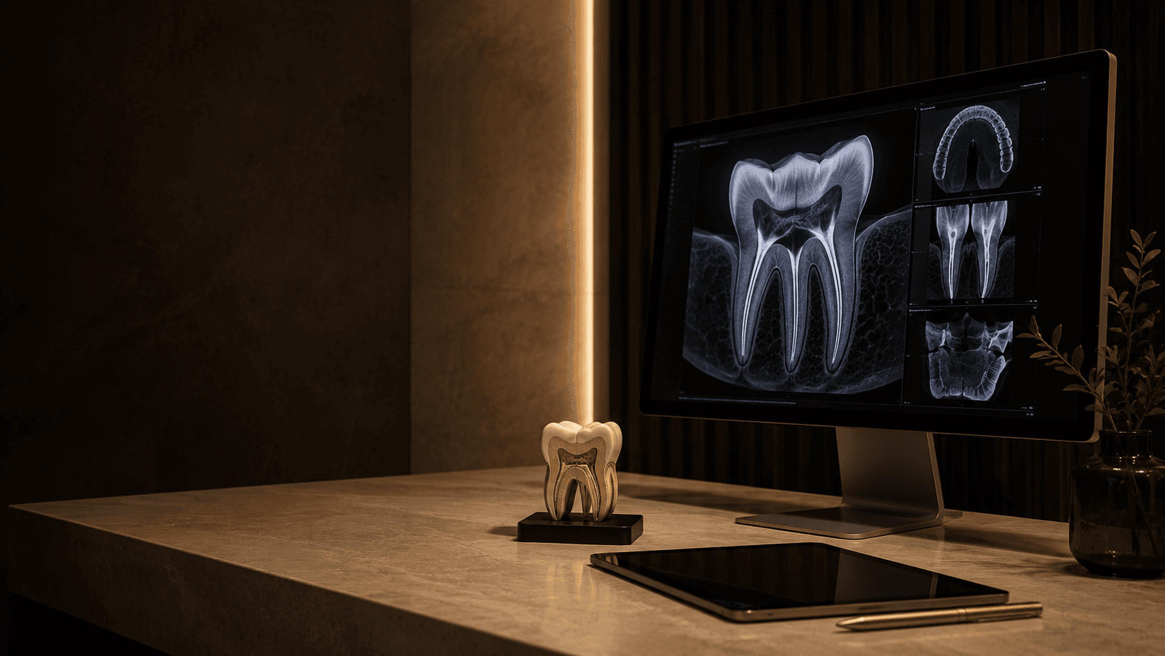
Every endodontic case at Madeel is performed under a Zeiss dental operating microscope at up to 25× magnification. The microscope is not optional kit — it is how the work is done. It lets us find canals that hide in molars, identify fractures before sealing, and remove old root-canal material cleanly during re-treatment. CBCT 3D imaging is used for complex and surgical cases. Most patients are surprised by how routine the appointment feels: local anesthetic, no pain during treatment, and back to normal eating once the numbness wears off.
Microscope at every step
Every root canal is performed under a Zeiss operating microscope at up to 25× magnification. Hidden canals, fine fractures and missed anatomy that a naked-eye procedure misses are visible — and treatable.
CBCT diagnosis
Complex and re-treatment cases are planned with 3D cone-beam imaging. We see the full root system, the surrounding bone and the source of infection before opening the tooth.
Single-visit where possible
Most microscopic root canals are completed in one appointment. The canal is cleaned, shaped, sealed and the tooth is prepared for its final restoration the same day.
Restored, not just treated
A treated tooth needs a proper seal to last. Our endodontic and restorative work runs together — post, core and crown planned from the start.
What to expect, step by step.
- 01
Diagnosis: clinical exam, digital X-ray, CBCT for complex cases.
- 02
Local anesthesia — the tooth and surrounding area are fully numbed.
- 03
Access opening through the crown of the tooth, under microscope.
- 04
Microscopic cleaning, shaping and disinfection of each canal.
- 05
Sealing of the canals with biocompatible filling material.
- 06
Final restoration — usually a crown — to protect the tooth long-term.
Why root canal beats extraction.
An implant or bridge can replace a missing tooth, but nothing matches a healthy natural root for bite, bone preservation and feel. As long as the tooth is restorable, saving it is the better long-term answer — and microscopic endodontics dramatically improves the odds.
- Saves the natural tooth instead of extraction and implant.
- Higher success rate than non-microscopic root canal.
- Most cases completed in a single visit.
- Pain-free under local anesthetic.
- Preserves jawbone and bite by keeping the natural root.
- Lower long-term cost than tooth replacement.
Not sure which treatment you need? Talk to a specialist on WhatsApp →
Common questions.
Direct answers to the questions patients ask before booking — pain, sedation, recovery, and the steps from consultation to result.
No. The tooth and surrounding gum are fully numbed with local anesthetic before any work starts. Most patients describe it as feeling similar to having a filling. Any pain you feel beforehand usually settles within a day of treatment.
Most microscopic root canals are completed in a single appointment of 60 to 90 minutes per tooth. Complex re-treatment or surgical cases may need a second visit.
For molars and pre-molars, yes — a root-treated back tooth is more prone to fracture and needs a crown to protect it. Front teeth with small access cavities can sometimes be restored with a bonded filling instead.
With a properly sealed restoration and routine dental care, a root-treated tooth can last decades. Long-term success depends more on the quality of the final crown and your hygiene than on the root canal itself.
Yes, modestly. The microscope, the time it takes to use it properly, and the higher success rate make it more costly than a conventional root canal. For most patients the trade-off is worth it — fewer re-treatments later.
The most common reasons are a canal that was missed during the original treatment, bacteria sealed inside, a crown that has started to leak, or a new crack in the tooth. Re-treatment under microscope addresses the underlying cause.
If the tooth is restorable, yes. A natural root preserves bone and bite in a way an implant cannot fully match. If the tooth is fractured below the gum or unrestorable, an implant is the better path.
Under microscope and ultrasonic tips, we can usually remove or bypass a separated instrument. CBCT lets us see exactly where the fragment is before we start, so we know going in whether retrieval is realistic.
The next step is a conversation.
Send a message, share what's going on, and we'll take it from there.
Prefer to call? +971 50 177 9179